This web page was produced as an assignment for Genetics 677, an undergraduate course at UW-Madison.
Introduction
Burkitt Lymphoma
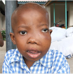
Figure 1. Swollen Jaw of eBL Infected Individual. Image taken from 50 years of Burkitt Lymphoma(4)
Burkitt Lymphoma (BL) is a rare form of cancer affecting the B-Cells of the immune system. 30-50% of lymphomas found in children are of the Burkitt variety as well as 1-2% of the lymphomas present in adults (3). There are three subtypes of BL: endemic BL (eBL), sporadic BL (sBL), and acquired immunodeficiency syndrome-related BL (AIDS-BL). eBL is predominantly seen in younger children with a "peak age incidence of 7 years" and affects a variety of organs and tissues (1). eBL is localized to Africa (4). The individual in Fig. 1 displays the age dependent attribute of a swollen jaw typically associated with eBL. sBL is often observed in children and young adults and is associated with the cases of BL not observed in Africa (4). Those inflicted with sBL exhibit irregular cell masses of the lower abdomen. AIDS-BL causes irregularities of the lymph nodes and bone marrow. Young adults most commonly show this subtype of BL (1). While BL occurs throughout the world, it is most prevalent in Central Africa as the endemic form(often in accordance with the Epstein-Barr Virus (EBV)) (2,3). The clinical symptoms of BL are a result of modifications to the MYC gene.
MYC Gene
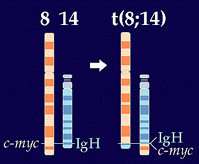
MYC is a protooncogene that is positioned on chromosome eight and is mapped to 8q24.12-q24.13. The protein that is translated from the MYC locus is a DNA-binding factor. DNA-binding factors are capable of activating or repressing the transcription of genes. The translated product of MYC controls cellular functions, in particular, cell growth and differentiation. MYC is also essential to DNA replication. Disruption of the MYC protooncogene results in a state of continual cell cycle without further differentiation, ultimately leading to tumor formation (1). A translocation between the c-myc gene and the immunoglobin (Ig) genes on chromosome 2, 14, or 22 is characteristic of the morphologies observed in BL (3). See Fig. 2. The translocations between MYC and the Ig genes affects the genes encoding for the kappa and lamba light chains as well as the heavy chain of Igs. With MYC placed under the control of the Ig locus, constitutive expression is observed in B-cells. The affected B-cells accumulate protein and eventually tumorigenesis occurs with uncontrollable cell growth and cell proliferation (7). Today, the cause of this translocation is still unknown.
Website Visitors
References:
1Wright, D. H. (1999). What Is Burkitt's Lymphoma and When Is It Endemic? Blood, 93(2), 758-759. Retrieved
from:http://bloodjournal.hematologylibrary.org
2NCBI Genes and Disease-Burkitt Lymphoma
3OMIM-Burkitt Lymphoma
450 Years of Burkitt Lymphoma
5WhosAmungUs
6PDB
7Thorley-Lawson, DA., Allday, MJ. (2008). The Curious case of the tumor virus: 50 years of Burkitt Lymphoma. Nature Reviews. 6:913-922
Contact Info: Deeter Neumann, [email protected], May 14, 2009
