This web page was produced as an assignment for Genetics 677, an undergraduate course at UW-Madison.
Phenotypes
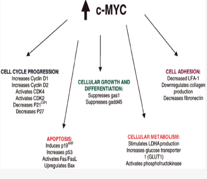
Due to MYC being an oncogene, many human cancers can be linked to an overexpression of MYC. The phenotypes of cells bearing overexpressed MYC proteins often result in "morphological abnormalties". These abnormalties can often be associated with differences in cell size, irregular control of the cell cycle, insensitivity to growth factors, incapable of differentiation, increased rates of apoptosis, and other genetic instabilities.(3) MYC has been shown to regulate many other target genes. With disruptions in MYC, signaling to these target genes is irregular and can often lead to other phenotypes that display cancerous properties. One of these target genes is involved in the WNT pathway and leads to irregular mammary cells and thus breast cancer.(4)
The figure on the right shows the functions in which MYC normally participates. Mutations arising in MYC can lead to the cellular phenotypes as noted above. Image taken from canceruptodate.com
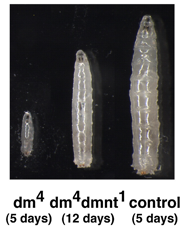
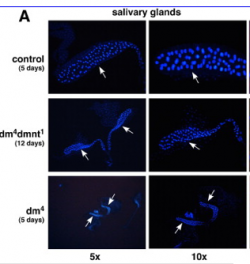
MYC Phenotype in Drosophila

Burkitt Lymphoma Phenotype
For an overview of the phenotypes associated with Burkitt Lymphoma please visit the following link:
Burkitt Lymphoma Overview Particularly the pathophysiology.
The image to the left is of a child suffering from the endemic form of Burkitt Lymphoma.(5)
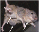
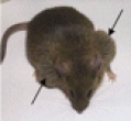
Mouse Model of Burkitt Lymphoma

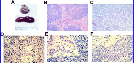
"Burkitt" Mice
Gene Ontology
The MYC protein can be found in all three of the gene ontology (GO) categories which include biological processes, molecular function, and cellular component.
Biological Processes
Some of the biological processes that MYC is involved with include transcription, cell proliferation, and cell cycle arrest.
Molecular Function
The molecular functions of MYC are DNA binding, protein binding, and transcription factor activity.
Cellular Component
As MYC is involved with DNA binding, protein binding, and processes like transcription and cell cycle arreast it is of no surprise that the only cellular component in which MYC is found is in the nucleus.
For a complete breakdown of the GO annotation please visit EMBL-EBI
References:
1EMBL-EBI
2Uniprot
3Rothermund et al. (2005). C-Myc–Independent Restoration of Multiple Phenotypes by Two C-Myc Target Genes with Overlapping Functions. Cancer Research, (65). 2097-2107. Retrieved from: http://cancerres.aacrjournals.org/cgi/content/full/65/6/2097
4Cowling et al. (2007). c-Myc Transforms Human Mammary Epithelial Cells through Repression of the Wntand SFRP1 Inhibitors DKK1. Molecular and Cellular Biology, (27)14. 5135-5146. Retrieved from: http://mcb.asm.org/cgi/content/full/27/14/5135?maxtoshow=&HITS=&hits=&RESULTFORMAT=&fulltext=MYC+AND+WNT+AND+Breast+cancer&andorexactfulltext=and&searchid=1&FIRSTINDEX=0&resourcetype=HWCIT
5canceruptodate.com
6emedicine
7Pierce, SB., Yost, C., and Andersen, S. (2008). Drosophila growth and development in absence of dMyc and dMnt. Developmental Biology. 315(2):303-316. Retrieved from: http://www.sciencedirect.com/science?_ob=ArticleURL&_udi=B6WDG-4RFJ4N1-1&_user=10&_rdoc=1&_fmt=&_orig=search&_sort=d&view=c&_acct=C000050221&_version=1&_urlVersion=0&_userid=10&md5=9b9a25effa5156ae728c4d67c7b2e772
8Boxer, LM., Wang, j. (2005). Regulatory elements in the immunoglobin heavy chain gene 3’-enhancers induce c-myc deregulation and lymphomagenesis in murine B cells. Journal of Biological Chemistry. 280(13). Retrieved from: http://www.jbc.org/cgi/content/full/280/13/12766
9Mice photos
10Affected Systems Table
Contact Info: Deeter Neumann, [email protected], May 14, 2009
